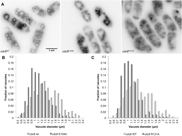
Fig. 4.
Analysis of vacuole fusion in cdc8E104A using the lipophilic dye, FM4-64. Cells were incubated for 45 min. in FM4-64, washed and transferred to medium as described in Materials and Methods. The images are of live cells after 60 min. (A) cdc8wt (SH30), cdc8E104A (SH41), cdc8R121A (SH39). (B) The size distribution of vacuoles in cdc8wt and cdc8E104A cells based on two independent experiments, each n>140. Mean±s.d.: cdc8wt, 1.4±0.4 µm; cdc8E104A, 1.1±0.3 µm (P=4.71×10⁻³¹). (C) The size distribution of vacuoles in cdc8wt and cdc8R121A cells in two independent experiments, each n>138. Mean±s.d.: cdc8wt, 1.3±0.3 µm; cdc8R121A, 1.1±0.2 µm (P=5.52×10−42). In both mutants the vesicles are smaller than in wildtype but greater in number. The vacuole size distributions in cdc8D16A.K30A, cdc8D131A, and cdc8E138A are indistinguishable from cdc8wt.

