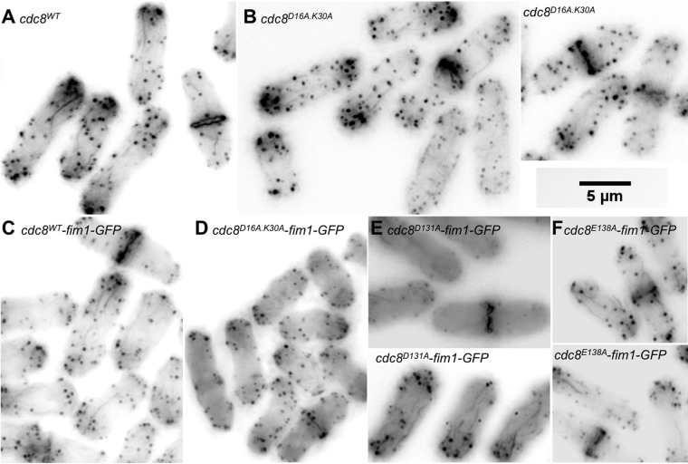
Fig. 5.
Actin cytoskeleton in wildtype and mutant cdc8 cells expressing GFP-fimbrin, illustrating synthetic negative effect of cdc8D16AK30A and fim1-GFP. All cells were fixed and stained with Alexa phallodin. Fim1-GFP fluorescence is weak following fixation. (A) cdc8wt cells (SH30). (B) cdc8D16AK30A cells (SH22). (C) cdc8wt fim1-GFP cells (SH77). (D) cdc8D16AK30A fim1-GFP cells (SH112). Note the absence of cables. (E) cdc8E131A fim1-GFP cells (SH74). (F) cdc8E138A fim1-GFP cells (SH75-A). The actin cytoskeleton appears normal in E and F.

