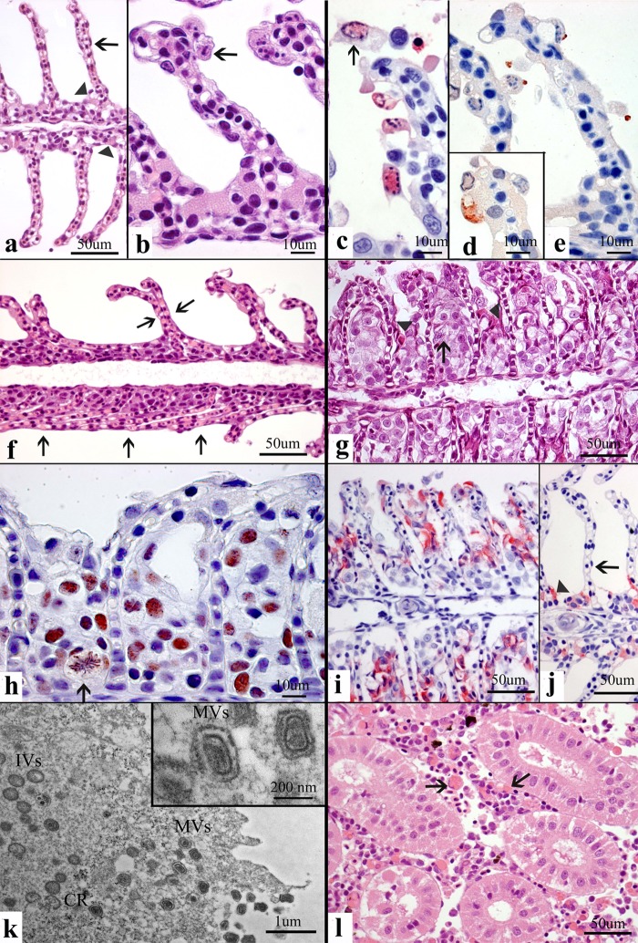
FIG 1.
Normal tissues and pathology in SGPV-infected Atlantic salmon. (a) A normal gill with thin lamellae (arrows) ensures efficient gas exchange. Chloride cells are present in normal numbers and at the normal location (arrowheads). (b and c) Detaching apoptotic cells with central clearing of chromatin (arrows) in the nuclear seen by H&E staining (b) and confirmed by red TUNEL staining (c). (d and e) IHC staining of poxvirus (brown) as cytoplasmic granules (d) and apical budding processes from apoptotic gill epithelial cells (e). (f) H&E staining of collapsed, adherent (arrows) thin lamellae losing apoptotic epithelial cells, creating an atelectasis-like condition hindering gas exchange. (g) H&E staining of proliferating (the arrow indicates metaphase), pale, foamy epithelial cells occluding the normally water-filled interlamellar space for gas exchange. Chloride cells are displaced and degenerated (arrowhead). (h) The lesion in panel g stained by IHC for PCNA showing brown nuclei, including proliferating cells in metaphase (arrow). (i and j) The lesion in panel g stained by IHC for chloride cells (red) that are displaced and enlarged (i) compared to the chloride cells in a normal gill (j). (k) TEM showing virus particles consistent with poxvirus in size and shape. Note the presence of crescents (CR), immature virions (IVs), and mature virions (MVs). (l) H&E staining of prominent hemophagocytosis (arrows) in the hematopoietic interrenal tissue. Methods included H&E staining (a, b, f, g, l); IHC staining for TUNEL (c), salmon gill poxvirus (d, e), PCNA (h), and chloride cells (i, j); and TEM (k).

