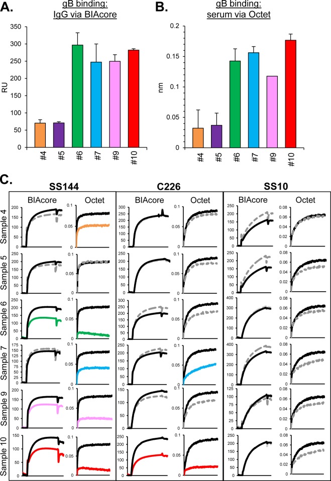
FIG 2.
Samples from HSV-infected humans, serum versus IgG. Antibody binding to gB was tested using purified IgG on a BIAcore 3000 biosensor (A) or using serum on an Octet RED96 system (B) as described in Materials and Methods. Relative binding units (RU) are indicated on the y axis. The averages of at least two experiments are shown; error bars indicate the standard errors. (C) Blocking of neutralizing anti-gB MAbs via human subject IgG (BIAcore) or sera (Octet). Binding curves for the association of test MAbs (SS144, C226, and SS10) to gB are shown. Black lines indicate MAb binding to gB that was not exposed to human subject samples (positive control). Gray dotted lines denote samples that block MAb binding less than 25% that of control. The curves reflect the extent to which Abs within the serum blocked MAb binding (steep curve, close to the black control curve = no MAb blocking; shallow curve = MAb blocking). The data using human IgG on the BIAcore for these six samples was first reported in Cairns et al. (35).

