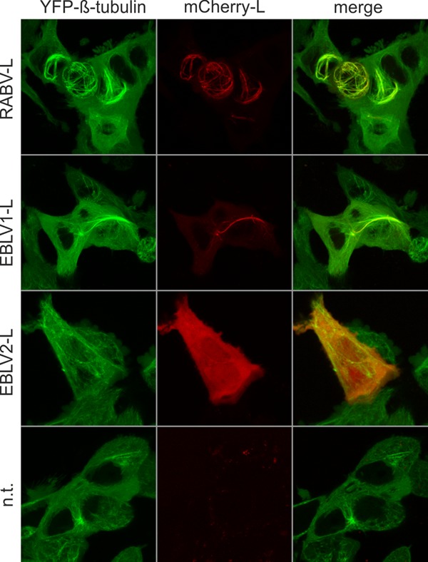
FIG 4.

Both RABV-L and EBLV1-L, but not EBLV2-L, accumulate at microtubules. Localization of mCherry-tagged L proteins of RABV (RABV-L), European bat lyssavirus type 1 L (EBLV1-L), and European bat lyssavirus type 2 L (EBLV2-L). Live confocal imaging was performed at 16 h posttransfection of expression plasmids into HEK-YFPtub cells. Not transfected (n.t.) cells were imaged as a negative control. All images are maximum projections of z-stacks acquired by live confocal imaging (optical slice, 0.772 μm; step size, 0.36 μm).
