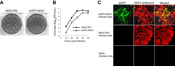
FIG 2.

Plaque morphology and growth kinetics of recombinant viruses. (A) Comparison of plaque sizes between AKAV-RG and eGFP-AKAV. (B) Growth curves for AKAV-RG and eGFP-AKAV in HmLu-1 cells. HmLu-1 cells were infected with eGFP-AKAV or AKAV-RG at an MOI of 0.01. The results are presented as the means ± SD (error bars) from three independent experiments. (C) Fluorescence microscopic analysis of cells infected with eGFP-AKAV. HmLu-1 cells were infected with eGFP-AKAV or AKAV-RG at an MOI of 0.01. (Left) eGFP fluorescence in cells infected with eGFP-AKAV or AKAV-RG or in mock-infected cells. (Middle) Infected cells that were incubated with an AKAV antiserum. (Right) Colocalization of AKAV antigens (red) and eGFP fluorescence (green).
