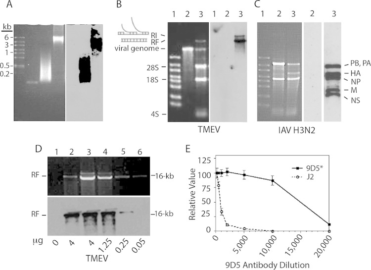
FIG 1.
Reactivity of the 9D5 MAb with dsRNA duplexes of various sizes and TMEV and influenza A virus RF RNAs. (A) (Left) Electrophoresis on a 1.7% agarose gel stained with ethidium bromide. The gel contained large molecular size ssRNA markers, a 47-mer RNA duplex, small-molecular-size (100- to 1,000-bp) poly(I·C), and large-molecular-size (1 to 8 kbp) poly(I·C) in the four lanes from left to right, respectively. (Right) Northwestern blot showing the reactivity of MAb 9D5 with 100-bp to 8-kbp poly(I·C). (B) (Left) Electrophoresis on a 1% agarose gel of in vitro-transcribed TMEV RNA and total RNA from TMEV-infected BHK-21 cells (6 h p.i.) stained with ethidium bromide. Lanes: 1, high-range ssRNA markers; 2, in vitro-transcribed TMEV genomic RNA; 3, total RNA from infected cells showing the TMEV positive-strand genome and dsRNA RF along with cellular RNAs. (Right) Northwestern blot of the membrane on the left showing the reactivity of RF and RI RNAs with 9D5 antibodies to dsRNA. (C) (Left) Electrophoresis on a 1% agarose gel of total RNA from uninfected and IAV-infected MDCK cells stained with ethidium bromide. Lanes: 1, ssRNA markers; 2, uninfected MDCK cells; 3, infected MDCK cells. (Middle) Northwestern blot of lane 2 from the panel on the left with no reactivity. (Left) Blot of the membrane of lane 3 from the panel on the left showing the reactivity of IAV genome RF segments. (D) Sensitivity of the 9D5 MAb for the detection of TMEV RF RNA from infected BHK-21 cells. (Top) A 1% agarose gel stained with ethidium bromide showing barely detectable TMEV RF RNA; (bottom) Northwestern blot showing the reactivity of TMEV RF RNA with as little as 50 ng of antibody. (E) Titration of MAb 9D5 (3.27 μg/μl) and MAb J2 (1.2 μg/μl) showing their relative reactivity with TMEV-infected M1-D cells by immunofluorescence intensity.

