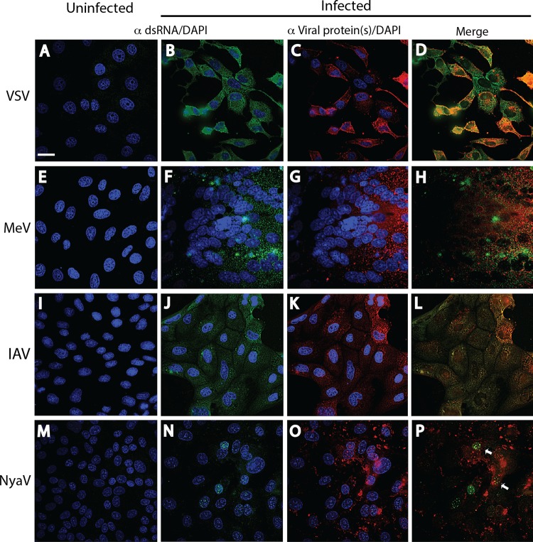
FIG 3.
Immunofluorescence analysis of dsRNA and virus proteins in negative-strand RNA virus infections. Uninfected (A, E, I, M) and infected (B, F, J, N) cell monolayers were stained with a 1:2,000 dilution of MAb 9D5 or polyclonal antibody 170A, and the other infected monolayers were stained with the specific antiviral antibodies (C, G, K, O). (A to D) Vero B6 cells infected or not infected with VSV. (A) Uninfected cells. (B) VSV-infected Vero B6 cells with punctate cytoplasmic staining (green) with MAb 9D5 (B). (C) VSV-infected cells stained with a 1:500 dilution of rabbit polyclonal antibodies to VSV G protein showing copious cytoplasmic staining (red). (D) Merged image of panels B and C. (E to H) Vero B6 cells infected or not infected with MeV. (E) Uninfected cells. (F) MeV-infected Vero B6 cells with punctate cytoplasmic staining with MAb 9D5. Note the large multinucleated giant cell. (G) MeV-infected cells were stained with human recombinant antibody to MeV NC protein and show diffuse cytoplasmic reactivity. (H) Merged image of panels F and G. (I to L) MDCK cells infected or not infected with IAV. (I) Uninfected cells. (J) IAV-infected MDCK cells revealing cytoplasmic staining (green) with MAb 9D5. (K) Cytoplasmic reactivity is seen with a 1:500 dilution of rabbit polyclonal antibody to IAV NC protein. (L) Merged image of panels J and K. (M to P) Vero B6 cells infected or not infected with NyaV. (M) Uninfected cells. (N) NyaV-infected Vero B6 cells revealing punctate areas of staining in the nucleus of infected cells. (O) Cytoplasmic reactivity with a 1:2,000 dilution of mouse antiserum to NyaV proteins is seen. (P) Merged image of panels N and O, with only a few of the stained nuclear dots in two cells showing colocalization (arrows). Bar, 10 μm (the magnification in panel A applies to all panels).

