FIG 5.

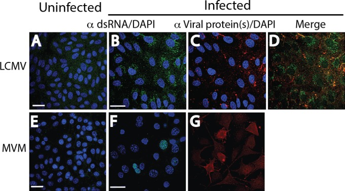
Reactivity of dsRNA and viral proteins in cells infected with an ambisense RNA virus (LCMV) and an ssDNA virus (MVM) by immunofluorescence analysis. Uninfected (A, E) and infected (B, F) cell monolayers were stained with a 1:2,000 dilution of MAb 9D5 to dsRNA, and infected monolayers were stained with the specific antiviral antibodies (C, G). (A to D) LCMV-infected and uninfected Vero B6 cells. (A) Uninfected cells. (B) LCMV-infected Vero B6 cells revealing punctate cytoplasmic staining (green) with MAb 9D5. (C) LCMV-infected cells stained with a 1:100 dilution of MAb to the LCMV NP protein showing cytoplasmic staining (red). (D) Merged image of panels B and C. (E to G) MVM-infected and uninfected mouse A9 cells. (E) Uninfected cells. (F) MVM-infected mouse A9 cells showing punctate areas of various diameters with staining with MAb 9D5 in the nucleus (green). (G) MVM-infected monolayers showed cytoplasmic staining with a 1:200 dilution of polyclonal antibody to MVM NS1/2 (no DAPI counterstaining) (red). The images in panels F and G were not from the same field. Bars, 10 μm (the magnification in the panels with bars applies to all photomicrographs).
