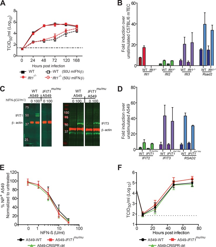
FIG 2.
Effects of human and mouse IFIT1 proteins on influenza A virus infection and replication. (A) Kinetics of IAV-Cal replication in WT and Ifit1−/− primary mTECs after infection at an MOI of 0.01, with or without IFN-β stimulation. The virus was harvested at 0, 24, 48, 72, 120, and 168 h postinfection, and viral yields were quantified by a TCID50 assay on MDCK cells. Data were pooled from two independent experiments performed in triplicate. (B) Expression of ISGs in WT and Ifit1−/− mTECs after IFN-β stimulation. mTECs from WT and Ifit1−/− mice were treated with 50 U/ml (dark colors) or 1,000 U/ml (light colors) of recombinant IFN-β or mock treated for 18 h before cellular RNA was extracted and used to quantify Ifit1, Ifit2, Ifit3, and Rsad2 gene expression by quantitative RT-PCR. Data were pooled from three independent experiments performed in duplicate. (C and D) Expression of ISGs in WT and IFIT1mu/mu A549 cells after IFN-β stimulation. (C) Western blots of human IFIT1, IFIT3, and β-actin on cell lysates from WT and IFIT1mu/mu A549 cells stimulated with 0 or 100 U/ml of human IFN-β for 18 h. (D) A549-WT and A549-IFIT1mu/mu cells were treated with 100 U/ml (dark colors) or 1,000 U/ml (light colors) of recombinant human IFN-β or mock treated for 18 h before cellular RNA was extracted and used to quantify IFIT2, IFIT3, and RSAD2 gene expression by quantitative RT-PCR. Data were pooled from two independent experiments performed in triplicate. (E and F) Kinetics of IAV-PR8 replication in WT and IFIT1mu/mu A549 cells. (E) Viral replication was measured in WT and IFIT1mu/mu A549 cells that were pretreated for 18 h with human IFN-β (hIFN-β). (F) WT and IFIT1mu/mu A549 cells were infected, and the virus titers in supernatants were determined at 1, 8, 24, 48, and 72 h postinfection. The data represent the means ± standard deviations (SD) for two independent experiments performed in triplicate.

