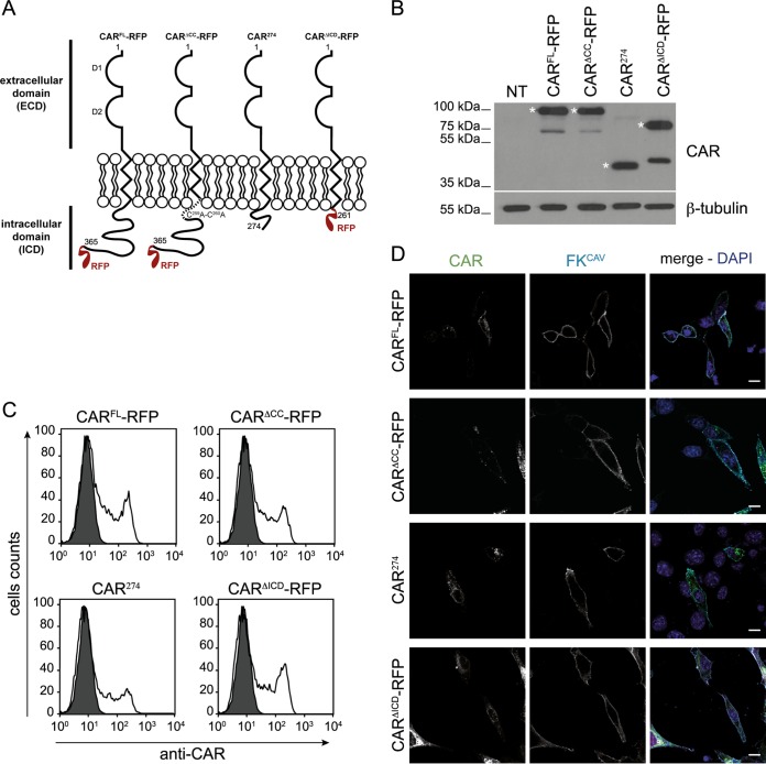
FIG 1.
Cell surface expression of CAR and its mutants in NIH 3T3 cells. (A) Schematic representation of constructs used in the study. The numbers indicate the amino acid residues, and the two Ig-like domains D1 and D2 are indicated. (B) Immunoblot analysis of CARFL-RFP, CARΔCC-RFP, CAR274, and CARΔICD-RFP expression. CAR was detected using an antibody against the ECD. Expected bands are indicated by an asterisk, and other bands correspond to CAR without the RFP tag. β-Tubulin was used as a loading control. Immunoblot analysis shows equal expression levels of the different DNA constructs in NIH 3T3 cells. NT, nontransfected. (C) Flow cytometry analysis of CAR cell surface levels. NIH 3T3 cells were transfected with plasmids expressing CAR constructs, and CAR cell surface expression was assayed by flow cytometry using an antibody against the CAR ECD on nonpermeabilized cells. (D) Confocal microscopy of CAR plasma membrane location using FKCAV. NIH 3T3 cells were transfected with the different constructs, and at 24 h posttransfection 1 ng/ml of FKCAV was added for 20 min on ice. Cells were fixed and stained for CAR (green) and for FKCAV (cyan). Scale bar, 15 μm.

