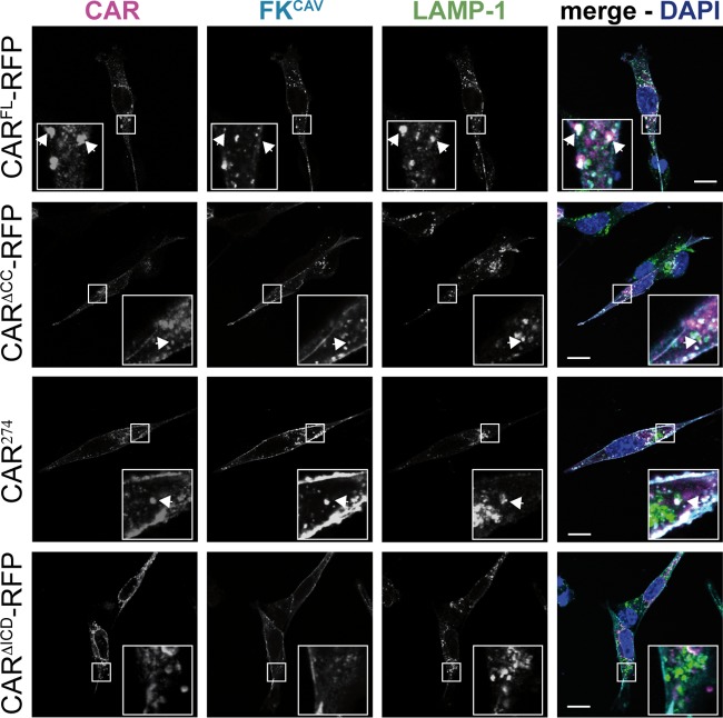
FIG 2.
CAR endocytosis triggered by FKCAV in NIH 3T3 cells. Confocal microscopy analyses were performed of FKCAV-mediated CAR endocytosis. NIH 3T3 cells were transfected with the different CAR constructs, and at 24 h posttransfection 1 ng/ml of FKCAV was added for 20 min on ice; samples were washed and incubated for 1 h at 37°C. Cells were labeled with anti-CAR against the ECD (magenta), anti-FKCAV (cyan), and anti-LAMP-1 (green) to visualize lysosomes. All constructs except CARΔICD-RFP allowed targeting of FKCAV to lysosomes. CAR lacking its ICD is not internalized upon FKCAV engagement. Insets show higher magnifications (∼3×) of internalized structures (white) containing CAR, FKCAV, and LAMP-1, identified by arrows. Scale bar, 10 μm.

