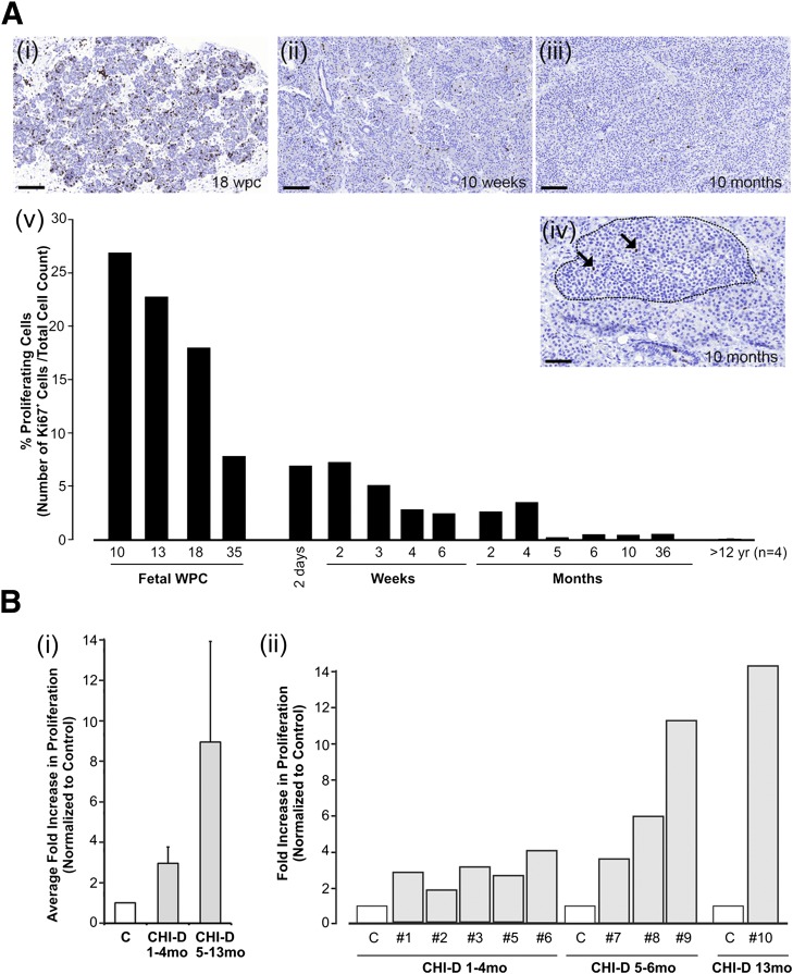
Figure 2.
Cell proliferation is increased in CHI-D tissue. A: Proliferation in human control pancreas from 10 wpc, during the first year after birth (weeks, months), and ≥12 years (yr). All data points were gathered from individual cases, except for the data associated with ≥12 years, which are averaged from four cases. Proliferation was assessed by high-density counting from a minimum of 20,000 cells, with Ki67+ cells expressed as a percentage of the total cell count. Ai–iv: Representative images from fetal tissue at 18 wpc and postnatal pancreas at 10 weeks and 10 months. Ki67+ cells are stained brown and are clearly seen in islets (arrows in panel Aiv). Scale bars represent 100 μm in Ai–iii and 50 μm in Aiv. B: Proliferation in CHI-D tissue. Bi: Average fold increase in Ki67 count expressed relative to age-matched controls from cases up to 4 months and the older ones. Bii: Individual data from the nine cases compared with age-matched controls demonstrate particularly higher proliferation rates at older ages. C, age-matched control; mo, months; #, patient identifier.

