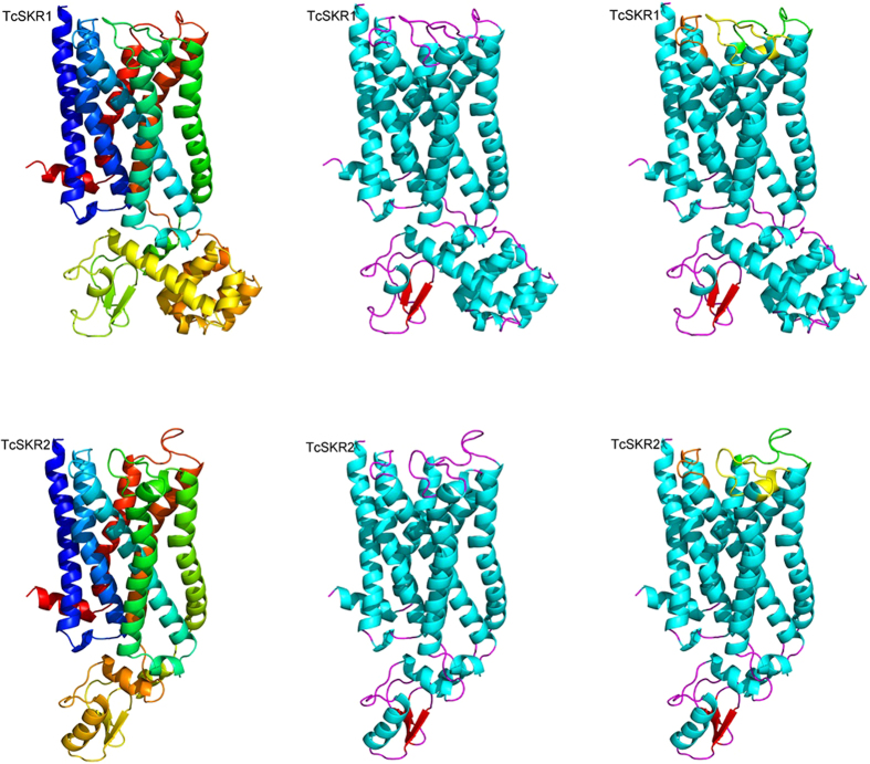
Figure 1. Cartoon diagram of Tribolium castaneum sulfakinin receptor 1 (TcSKR1) and T. castaneum sulfakinin receptor 2 (TcSKR2).
The seven transmembrane α-helices building the three-dimensional fold of the proteins are differently colored from blue (N-terminus) to red (C-terminus) (upper and lower on the left) by secondary structure (upper and lower in the middle) and with extracellular loops (ECL) colored differently (upper and lower on the right). The ECL 1 is colored in orange, ECL 2 in yellow and ECL 3 in green. The N-terminus is colored in blue while C-terminus red.

