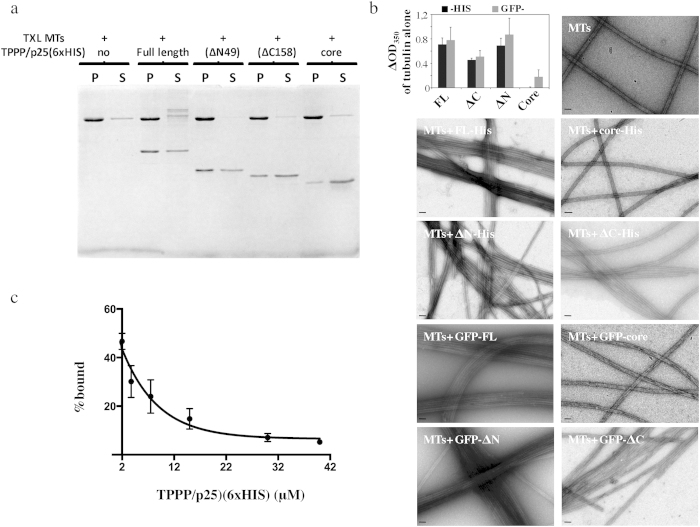
Figure 3. Microtubule binding properties of TPPP/p25.
(a) Taxol stabilized microtubules (2 μM) were incubated in the presence of 2 μM of each of the different TPPP/p25 fragments (FL, ΔN, ΔC and core) for 15 min before centrifugation. Coomassie-stained gel of microtubule bound and unbound TPPP/ p25 fragments present in the pellets and in the supernatants, respectively. (b) Bar graph showing the changes in turbidity of tubulin solutions (15 μM) in the presence of stoichiometric amounts of the different TPPP/ p25 fragments. The OD350 of control tubulin solutions at mass steady state were subtracted from the tubulin solutions assembled in the presence of the different TPPP/ p25 fragments tagged with either 6×HIS at their C-termini or GFP at the N-termini. Data were compared using the Mann Whitney test (p values for the HIS- vs the GFP- tagged fragments were 1, 0,666, 0,667 and 0,333 for FL, ΔN, ΔC, and core respectively; p values for the FL, or ΔN, or ΔC fragments compared to the core were 0,028, 0,2 and 0,57, respectively; n = 4). EM images of taxol stabilized microtubules (2 μM) incubated with bacterially expressed and then purified C-terminal His- or GFP-tagged TPPP/p25 fragments at equimolar ratios (scale bars = 50 nm). (c) His tagged TPPP/p25 competes with GFP-tagged TPPP/p25 for MT binding. Taxol stabilized MTs were first co-incubated with 2 μM of 6×HIS-FL and 2 μM GFP-FL-TPPP/p25 and then with increasing concentrations of 6×HIS-FL. Each data point represents the mean ± SD from three independent experiments.

