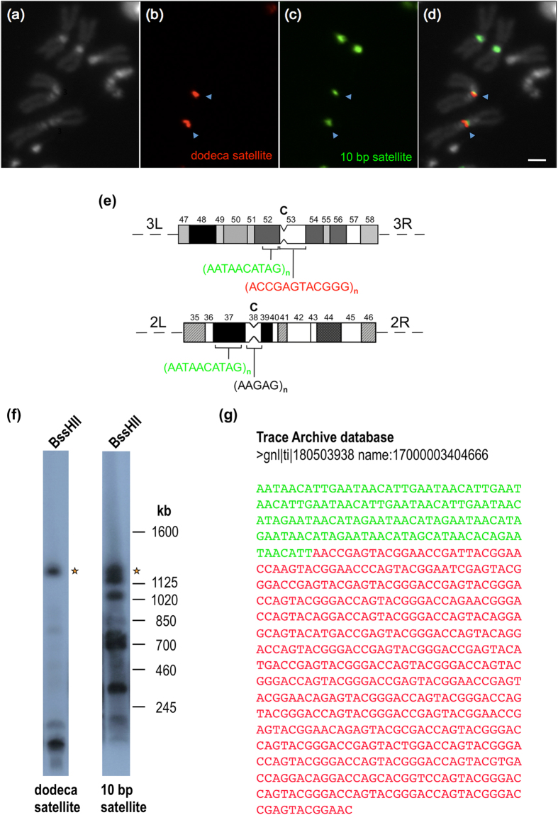
Figure 2. The 10 bp satellite DNA localizes on the third chromosome at h52p instead of h48.
(a) Metaphase chromosomes counterstained with DAPI. (b) Hybridization signals from a dodeca satellite probe (in red). (c) Hybridization signals from a 10 bp satellite probe (in green). (d) Hybridization signals superimposed with DAPI-stained chromosomes. The Scale bar is 2 μm. (e) Diagram representing the heterochromatic regions39 of chromosomes 2 (regions 35–46) and 3 (regions 47–58) showing the localization of the 10 bp (in green) and dodeca (in red) satellites. The position of the centromeres (C) is indicated. (f) High molecular weight DNA from red e embryos was digested with BssHII, electrophoresed through a 1% (w/v) agarose gel in a “Waltzer” apparatus at 150 V for 24 h with a 130 s pulse time, blotted onto a nylon filter and hybridized successively with the dodeca satellite probe pBK6E218 at 68 °C and with the 10 bp satellite probe 5′-AATAACATAGAATAACATAGAATAACATAGAATAACATAGAATAACATAG-3′ at 50 °C. The asterisks indicate a 1.2 Mb fragment that hybridizes with both probes. (g) DNA sequence showing a junction between 10 bp satellite repeats (in green) and dodeca satellite repeats (in red).

