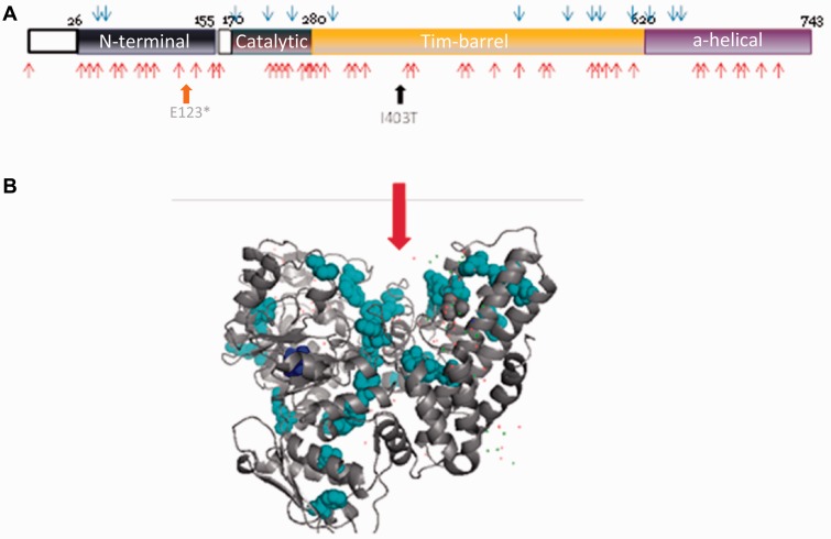
Figure 3.
Location of mutations within the NAGLU protein. (A) Linear representation of the four domains (N-terminal, catalytic, Tim-barrel, and alpha-helical) of the NAGLU protein showing the position of the p.Glu123* variant in the N-terminal domain and the p.Ile403Thr variant in the Tim-barrel domain. Blue arrows represent the position of mutations associated with attenuated phenotypes and red arrows the ones causing severe phenotypes. (B) 3D structure of the human NAGLU protein showing the p.Ile403Thr mutation (dark blue) and severe mutations clusters (light blue). The red arrow indicates the catalytic site.

