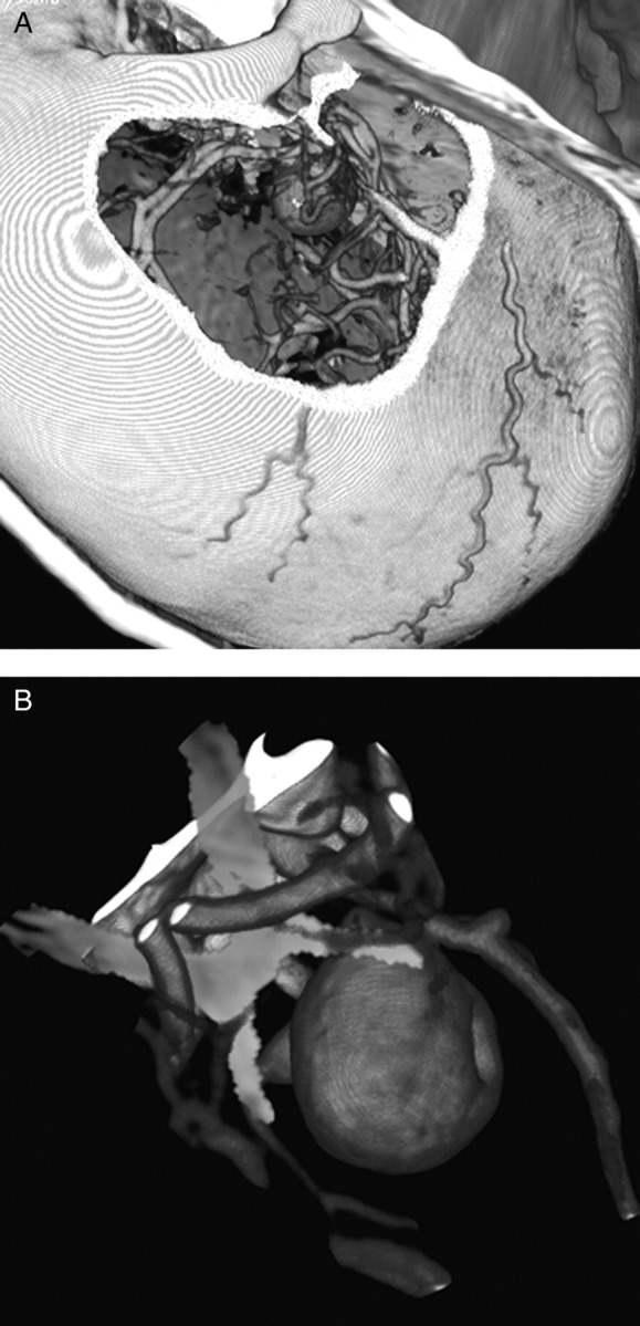
Figure 2.

(A). The CTA volumetric images were windowed for viewing the vasculature and skull anatomy. (B) MRI volumes were windowed for an optimum view of parenchymal anatomy, cranial nerves and cisternal spaces, here allowing for the rendering of the optic chiasm (in yellow).
