
Fig. 3.
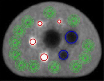
Position of the regions of interest (ROIs) placed over the active spheres (red), nonradioactive spheres (blue) and the phantom background (green) of the reconstructed PET images, which are used for analysis
Official websites use .gov
A
.gov website belongs to an official
government organization in the United States.
Secure .gov websites use HTTPS
A lock (
) or https:// means you've safely
connected to the .gov website. Share sensitive
information only on official, secure websites.
Position of the regions of interest (ROIs) placed over the active spheres (red), nonradioactive spheres (blue) and the phantom background (green) of the reconstructed PET images, which are used for analysis