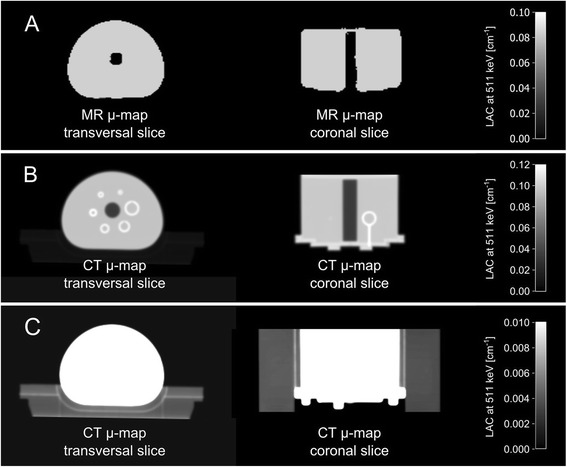
Fig. 4.

a MR-based μ-map in transversal and coronal orientation only contains discrete attenuation values for water and air, and therefore only corrects for photon attenuation caused by the water content of the phantom and not by the phantom housing materials, as these materials cannot be detected with standard MR imaging. b, c CT-based μ-map contains continuous attenuation values including μ-values for the phantom housing, glass spheres, and styrofoam block used as phantom holder (displayed in c). b and c visualize the same content, however windowing properties were adjusted individually in order to visualize either the phantom content (b) or the styrofoam holder, on which the phantom is placed (c)
