Figure 5.
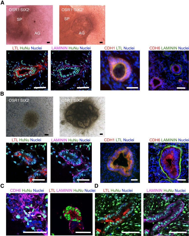
Developmental potential of OSR1+SIX2+ renal progenitors to differentiate into renal lineage cells or tissues. (A): The presented macroscopic views show the aggregates of OSR1−SIX2− cells on culture day 5 (upper left) and OSR1+SIX2+ cells on day 7 (upper right) cocultured with E11.5 mouse spinal cord tissue in organ cultures. Lower panels: Immunostaining images of the histological sections of the OSR1+SIX2+ cell aggregates after 7 days of coculture with spinal cord tissue. LTL is a marker of the proximal renal tubules; LAMININ, a marker of the polarized epithelia; CDH1, an epithelial marker; CDH6, an early proximal tubule marker. (B): Coculture experiments with NIH3T3 fibroblasts expressing Wnt4. Macroscopic views of the aggregates of OSR1−SIX2− cells on day 5 (upper left) and OSR1+SIX2+ cells on day 7 (upper right) and immunostaining images of the histological sections of the OSR1+SIX2+ cell aggregates after 7 days of coculture (lower). Note that the lower left two panels in (A) and (B) are of the same sections. (C): Immunostaining images of the organ culture of the OSR1+SIX2+ cells and E11.5 metanephric cells. (D): Immunostaining images of the OSR1+SIX2+ cell aggregates transplanted into the epididymal fat pads of the immunodeficient mice (NOD. CB17-Prkdcscid/J) after 30 days of transplantation. Scale bars = 50 μm. Abbreviations: AG, OSR1−SIX2− or OSR1+SIX2+ cell aggregate; HuNu, human nuclei; LTL, Lotus tetragonolobus lectin; SP, spinal cord.

