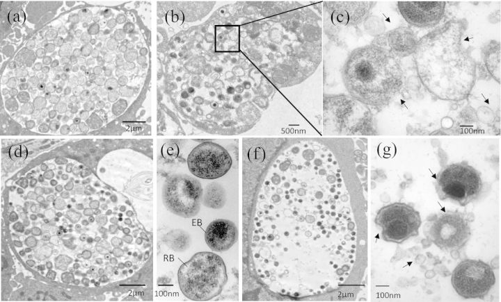
Figure 5.
Altered ultrastructure of chlamydial organisms in HeLa 229 cells induced by Andro. (a–c) Representative micrographs of infected cells fixed at 28 h pi. Cells unexposed (a) or exposed (b and c) to Andro (30 μM). An enlarged image from the box in (b) is shown in (c). (d–g) Representative micrographs of infected cells fixed at 36 h pi. Cells unexposed (d and e) or exposed (f and g) to Andro (30 μM). Note: deformed chlamydial organisms were only observed in C. trachomatis-infected cells exposed to Andro as indicated in (b), (c), (f) and (g). Arrows indicate irregular C. trachomatis membranes and MV accumulation induced by Andro. Scale bar for (a), (d) and (f) = 2 μm, for (b) = 500 μm, and for (c), (e) and (g) = 100 nm.

