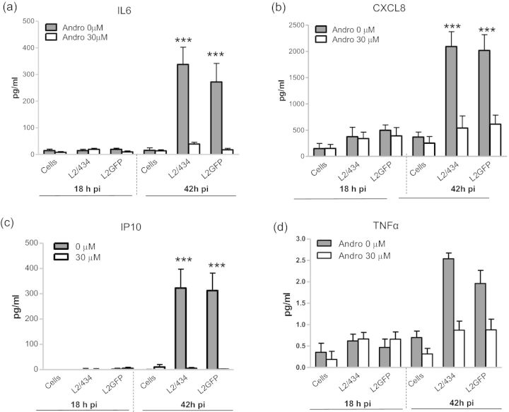
Figure 6.
Suppression of proinflammatory cytokine secretion from C. trachomatis-infected epithelial cells by Andro. (a) IL6; (b) CXCL8; (c) IP10; (d) TNFα. HeLa 229 cells were infected with EBs of L2/434 or L2GFP, followed by the immediate addition of Andro (30 μM) to the culture. Culture supernatants were collected at 18 and 42 h pi and measured for levels of cytokines using cytometric bead assays. Data bars show the mean ± SD (pg/ml). ***P < 0.001, compared with the mock-infected cell control using one-way ANOVA and Bonferroni's test. No significant difference in TNFα production was observed between L2/434- or L2GFP-infected cells and the mock-infected control (P > 0.05).

