Figure 3.

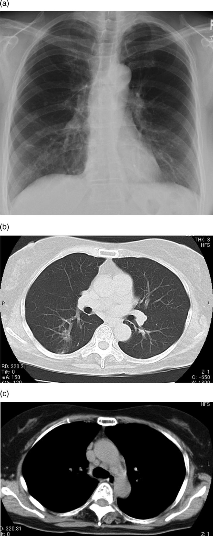
(A) Plain chest radiograph showing improvements of bilateral hilar lymphadenopathy and nodular shadows. (B) Chest CT showing decreases of the size of small nodules. (c) Hilar and mediastinal lymphadenopathy showing decreases in size.

(A) Plain chest radiograph showing improvements of bilateral hilar lymphadenopathy and nodular shadows. (B) Chest CT showing decreases of the size of small nodules. (c) Hilar and mediastinal lymphadenopathy showing decreases in size.