Figure 1.
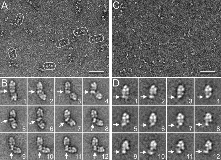
EM analysis of FVIII in complex with dimeric and monomeric D′D3. (A) Representative raw image of dimeric FVIII-D′D3 in negative stain. Some of the complexes are circled. The scale bar represents 50 nm. (B) Selected class averages of dimeric FVIII-D′D3 complex illustrating that the angle between the 2 FVIII molecules varies greatly. White arrows indicate the density representing the dimeric D′D3 domain. The side length of individual panels is 75.1 nm. (C) Representative raw image of monomeric FVIII-D′D3 in negative stain. The scale bar represents 50 nm. (D) Selected class averages of monomeric FVIII-D′D3 complex illustrating that structural heterogeneity persists in this complex. White arrows indicate the density representing the monomeric D′D3 domain. The side length of individual panels is 33.4 nm.

