Figure 2.
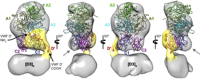
The D′ domain interfaces primarily with the FVIII C1 domain. 3D reconstruction of the murine [D′D3]2-FVIII ternary complex (determined from 962 projections) reveals the D′ density (suggested by the yellow shell) extending from the [D3]2 core and enmeshed with the region corresponding to the FVIII C domains. The structure of FVIII14 (Protein Data Bank: 2R7E) fits the EM density in a unique orientation (A1, olive; A2, green; A3, cyan; C1, blue; C2, purple). The suboptimal agreement between the EM envelope and the position of the C domains suggests that they likely undergo conformational changes upon interaction with VWF D′. Modeling of the VWF D′ domain (Protein Data Bank: 2MHP, red) within the corresponding EM density suggests that it interacts primarily with the FVIII C1 domain, although more limited interactions are possible with C2 and A3. The N- and C-termini of the VWF D′ domain are represented by spheres and indicated by black arrows.

