
Figure 1.
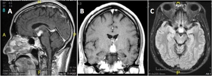
(A) Sagittal T1 post contrast. (B) Coronal T1 post contrast. (C) Axial T2 Flair. T1 and T2-weighted MRI images depicting hypothalamic hyperintensities. T2 images show hyperintensity in the anterior hypothalamus, with signal abnormalities lining the mildly expanded anterior third ventricle. No brainstem involvement appeared in MR images at that time, nor were there any appreciable lesions of the locus coeruleus or raphe nuclei.