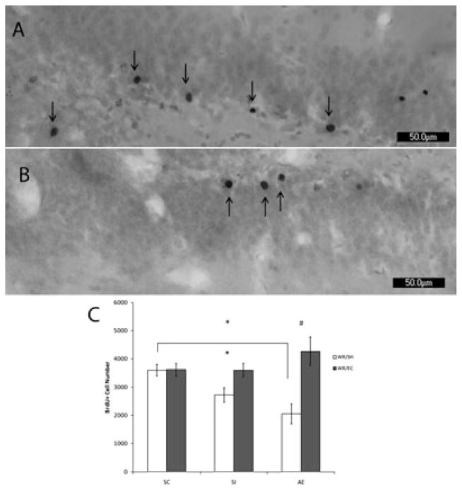
Figure 3.
PD72 analysis of BrdU labeling using light microscope. Images of BrdU+ labeled cells recognized by reaction with diaminobenzidine and counterstained with Pyronin Y. Images taken at 20×. (A) is taken from an AE animal exposed to WR/EC. (B) is taken from an AE animal exposed to WR/SH. (C) demonstrates that the number of BrdU+ cells is significantly decreased in AE animals compared to SC when exposed to standard housing after running (p<0.05). Exposure to environmental complexity significantly increases the number of surviving new cells in both the SI (p < 0.05) and the AE (p < 0.01) groups compared to social housed littermates. All values represent mean +/− SEM. * p < 0.05; # p < 0.01.

