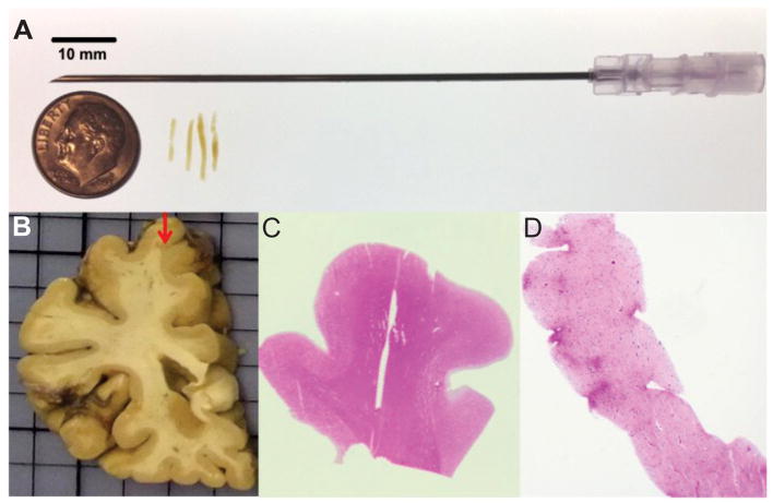
Figure 1.
Needle core biopsy of the frontal lobe cortex of autopsied subjects. (A) Four needle cores were collected with an 18-gauge needle from fixed tissue of deceased subjects with different neuropathologic diagnoses. (B) The cores were collected from the crest of the superior frontal gyrus at the level of the head of the caudate nucleus. (C, D) All the cores and the source blocks used for biopsy were embedded in paraffin and sectioned. All cores and source blocks were stained with hematoxylin and eosin to confirm the presence of gray and white matter. Core image in (D) was taken at a 20x magnification.

