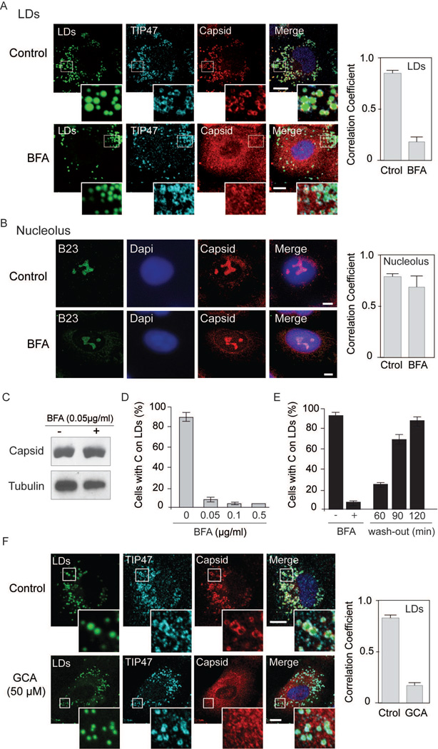
Figure 2.
Brefeldin A blocks C localization on LDs in DENV infected cells. A. Immunofluorescence showing the effect of BFA on C associated to LDs. Specific antibodies against C and TIP47 were used in infected cells as indicated on the top. LDs were stained with BODIPY. Representative confocal images are shown for each case in the absence or presence of 0.05 µg/ml of BFA. Bar, 5 µm. Quantification of the percentage of C that co-localizes with TIP47 (mean +/− standard deviation) was performed using ImajeJ (JACoP plug-in), Manders’ coefficient is shown. B. Immunofluorescence showing the effect of BFA on C accumulation in nucleolus using methanol as fixation method. Representative confocal images using antibodies against C and B23 (marker of nucleolus) are shown in the absence or presence of 0.05 µg/ml of BFA. Bar, 5 µm .Quantification of the percentage of C that co-localizes with B23 (mean +/− standard deviation) was performed using ImageJ (JACoP plug-in), Manders’ coefficient is shown. C. Total amount of C in cells treated or not with BFA. Cell extracts were used for immunoblots with specific anti-C antibodies. D. Effect of different concentrations of BFA on C association to LDs. Confluent A549 infected cells were treated at 8 h post-infection with 0, 0.05, 0.1 or 0.5 µg/ml of BFA. At 24 h post-infection cells were immunostained with anti-TIP47 and anti-C antibodies and the percentage of cells with C on LDs was determined. About 200 cells were scored; bars indicate the standard error of the mean. E. Confluent A549 infected cells were treated 8 h with BFA and then the BFA was removed. At 60, 90 and 120 min after washing, cells were fixed and stained. The percentage of cells with C on LDs is shown for each time. About 200 cells were scored in duplicates. Bars indicate the standard error of the mean. F. Golgicide A impairs C localization on LDs. Immunofluorescence showing the effect of 50 µM of Golgicide A on C association to LDs in A549 cells. Representative confocal images of cells treated or not with the drug (Control or GCA) are shown stained with BODIPY for LDs, or labeled with anti-TIP47 and anti-Capsid as indicated for each case. Bar, 10 µm. Right panel, quantification of the percentage of capsid that co-localizes with TIP47 (mean +/− standard deviation), Manders’ coefficient is shown.

