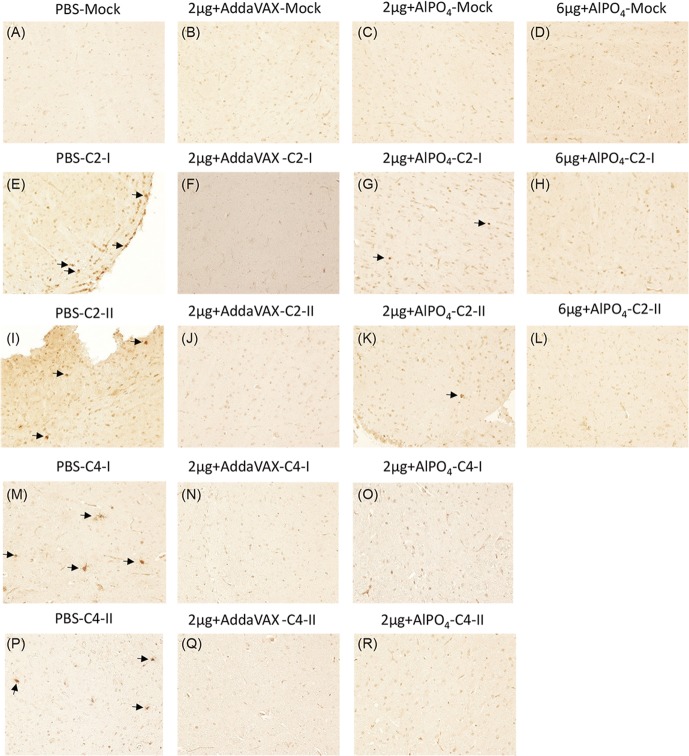
Fig 8. In situ detection of EV71 distribution in the brain stems of Tg mice after challenge.
The uninfected Tg mice either pre-treated with PBS or vaccines were used as the negative control, and the images of immunohistochemistry (IHC) staining with the Mab979 antibody are shown in A-D. The PBS- and vaccine-treated Tg mice infected with the EV71 C2 or C4 strains were sacrificed on day 6 post-infection, and the waxed sections of brain stem stained by IHC are representatively shown twice in E-R as labeled. All pictures were taken at 200X magnification. Viral particles in the sections are indicated with arrows.

