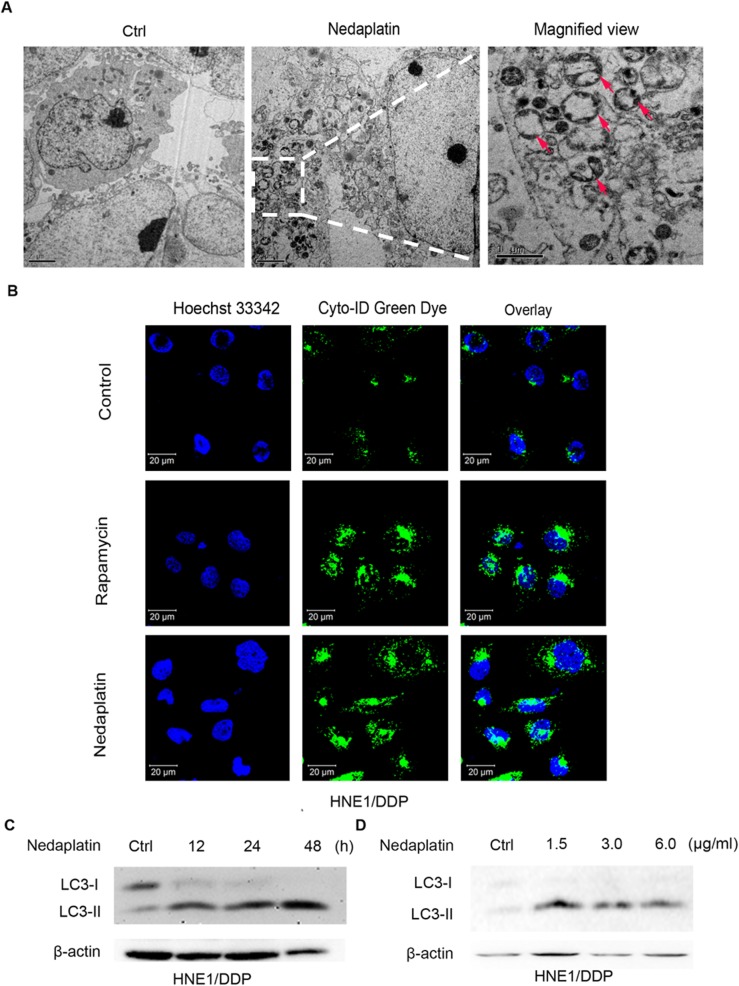
Fig 2. Autophagy was induced by nedaplatin in HNE1/DDP cells.
(A) Nedaplatin induced formation of autophagosomes. HNE1/DDP cells were either untreated or treated with 6.0 μg/ml of nedaplatin for 48 h. A magnified view of the electron photomicrograph shows a characteristic autophagosome. (B) HNE1/DDP cells were treated with 6.0 μg /ml of nedaplatin for 24 h or with 500 nM of rapamycin for 4 h, and then the cells were stained with Cyto-ID Green autophagy dye and analyzed by confocal microscopy. (C) Immunoblot analysis of LC3-I/II levels. HNE1/DDP cells were treated with 6.0 μg/ml nedaplatin for 12, 24, and 48 h. (D) Immunoblot analysis of LC3-I/II levels. HNE1/DDP cells were treated with nedaplatin for 48 h at 0, 1.5, 3.0 and 6.0 μg/ml.

