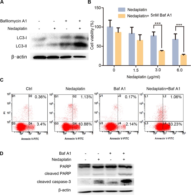
Fig 3. Inhibition of autophagy enhanced nedaplatin-induced apoptosis and suppression of cell growth in HNE1/DDP cells.
(A) HNE1/DDP cells were incubated with 6.0 μg/ml nedaplatin for 48 h, in the presence or absence of Baf A1 (5 nM) for 48 h, and the levels of LC3-I/II were detected by western blot. (B)HNE1/DDP cells were untreated or treated with nedaplatin at indicated concentrations in the absence or presence of Baf A1 (5 nM) for 48h. The cell viability was determined by MTT assay at the wavelength of 570 nm. Data are mean ± SD from five independent experiments. ***p<0.001 compared to nedaplatin only. (C) HNE1/DDP cells were treated with 6.0 μg/ml with or without Baf A1 (5 nM) for 48 h and then analyzed by FACS after PI and FITC-annexin V staining. (D) HNE1/DDP cells were incubated with or without 6.0 μg/ml of nedaplatin in the presence or absence of the autophagy inhibitors Baf A1 for 48 h. The whole protein was extracted, and PARP, cleaved PARP and cleaved caspase 3 were analyzed by Western blot.

