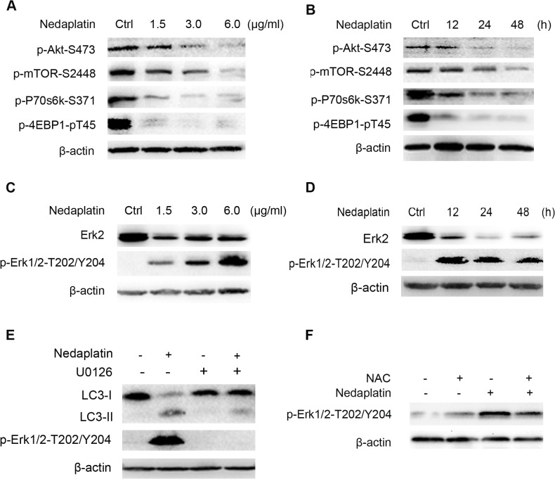
Fig 5. Akt/mTOR and ERK signaling pathways were involved in nedaplatin-induced autophagy in HNE1/DDP cells.
(A) HNE1/DDP cells were treated with different concentrations of nedaplatin for 48 h. Levels of pAkt, pmTOR, pP70S6K, and p4E-BP1 were detected by western blot. (B) HNE1/DDP cells were treated with 6.0 μg/ml nedaplatin as indicated times. Levels of pAkt, pmTOR, pP70S6K, and p4E-BP1 were detected by western blot. (C) HNE1/DDP cells were treated with different concentrations of nedaplatin for 48 h. Protein extracts were analyzed using Erk1/2 and phospho-Erk1/2 (Thr202/Tyr204) by western blot. (D) HNE1/DDP cells were treated with 6.0 μg/ml nedaplatin as indicated times. Protein extracts were analyzed using Erk1/2 and phospho-Erk1/2(Thr202/Tyr204) by western blot. (E) HNE1/DDP cells were treated with 6.0 μg/ml nedaplatin for 48 h with or without the pretreatment of U0126 (20 μM) for 2h. Protein extracts were examined by Western blot using LC3 I/II and phospho-Erk1/2 (Thr202/Tyr204) antibodies. (F) The phospho-Erk 1/2 (Thr202/Tyr204) levels were examined by western blot after the nedaplatin treatment with or without of NAC (10 mM) for 48 h.

