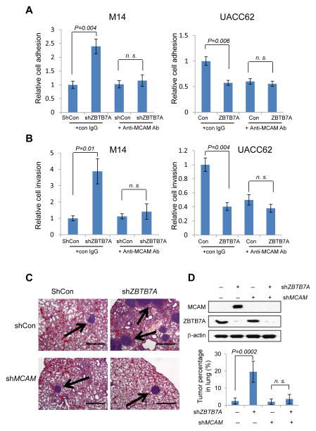
Figure 6.
ZBTB7A suppresses melanoma cell adhesion, invasion and in vivo metastasis by repressing MCAM. A, ZBTB7A was stably knocked down using lentiviral shRNA in M14 melanoma cell line that expresses a relatively high level of ZBTB7A. The cells were subject to a HUVEC cell adhesion assay in the absence or presence of anti-MCAM antibody (Left panel). Retrovirus mediated ZBTB7A overexpression in UACC62 melanoma cells that express a relatively low level of ZBTB7A. The cells were similarly subject to HUVEC cell adhesion assay. Data are mean ± s.d. of three independent experiments. B, the ZBTB7A stable knockdown M14 cells as in A were assessed for cell invasion through matrigel, again in the absence or presence of anti-MCAM antibody (Left panel). ZBTB7A overexpressing UACC62 cells were similarly assessed for cell invasion through matrigel (Right panel). Data are mean ± s.d. of three independent experiments. C, M14 melanoma cells stably expressing ShCon, shZBTB7A or shMCAM were generated and the expression levels of MCAM and ZBTB7A were analyzed by Western blot (D). The cells were injected into nude mice through tail vein. The animals were harvested 60 days later and lungs were collected for H&E staining. Representative H&E images of nude mice lung are shown. Bar, 500μm. Arrows indicate the lung nodules formed by melanoma cells. The tumor nodules were quantified and presented as percentage in each lung. The numbers represent mean ± s.d from 5 mice/group (D).

