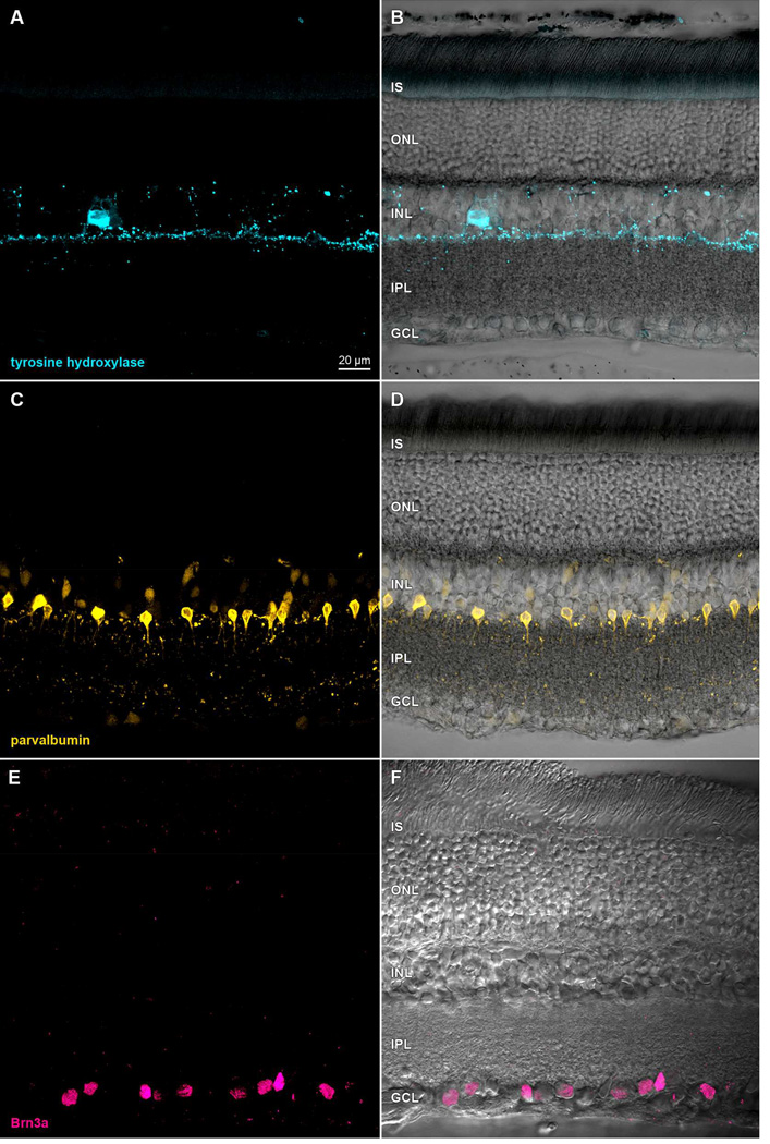
Figure 1.
Immunoreactivity in formaldehyde-fixed rat retina. Transretinal vibratome sections incubated in (A) anti-tyrosine hydroxylase primary antibody and Alexa Fluor 488- conjugated anti-mouse secondary antibody, (C) anti-parvalbumin primary antibody and Alexa Fluor 488-conjugated anti-mouse secondary antibody, or (E) anti-Brn3a primary antibody and DyLight 549-conjugated anti-goat secondary antibody. Paired panels show single optical sections (A,C,E) of fields imaged under epifluorescence illumination on a laser scanning confocal microscope, and after merging these with the same fields under differential interference contrast optics (B,D,F). Acronyms positioned at the inner segment (IS), outer nuclear (ONL), inner nuclear (INL), inner plexiform (IPL), and ganglion cell (GCL) layers. Scale bar in (A) is 20 µm and applies to (A–F).

