Figure 2.
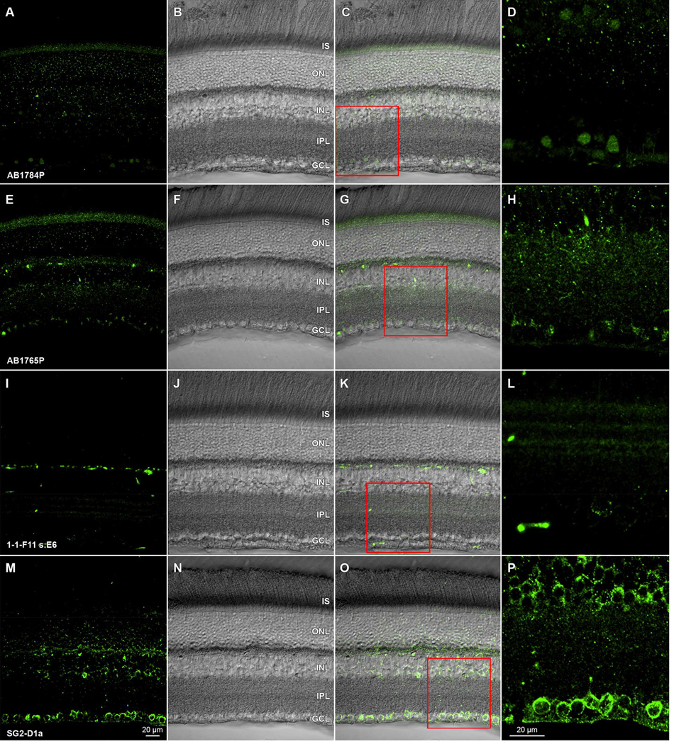
Antibody-specific immunoreactivity effects of formaldehyde fixation on antidopamine D1 receptor antibodies in rat retina. Transretinal vibratome sections from a single retina incubated in (A) rabbit polyclonal AB1784P, (E) rabbit polyclonal AB1765P, (I) rat monoclonal 1-1-F11 s.E6, or (M) mouse monoclonal SG2-D1a. All preparations are visualized with Alexa Fluor 488-conjugated species-specific secondary antibodies. Paired panels show single optical sections (A,E,I,M) of fields imaged under epifluorescence illumination, the same fields under under differential interference optics (B, F, J, N), and merges of these in (C,G,K,O). High magnification details (D,H,L,P) of the ganglion cells layer, inner plexiform layer, and proximal inner nuclear layer are shown, and correspond to the red square regions of (C,G,K,O), respectively. Acronyms positioned at retinal layer levels as in Fig. 1. Scale bar in (M) is 20 µm and applies to (A–C, E–G, I–K, M–O). Scale bar in (P) is 20 µm and applies to (D,H,L,P). Lack of detectable GCL immunopositivity in A–L contrasts with GCL immunopositivity in M–P and with ganglion cell dopamine-sensitivity (Hayashida et al., 2009; Ogata et al., 2012).

