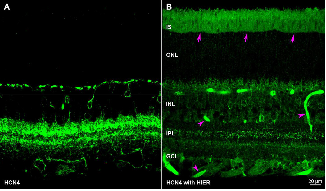
Figure 3.
Artifactual autofluorescence after heat induced epitope retrieval (HIER). Transretinal vibratome sections incubated in anti-HCN4 primary antibody and Alexa Fluor 488-conjugated anti-mouse secondary antibody. Paired panels show z-stacks of 5 optical sections under epifluorescence illumination. The fluorescence pattern in untreated retina (A) differs from HIER-treated tissue (B) in that HIER introduces extraneous staining in the photoreceptor inner and outer segments (arrows) as well as blood vessels (arrowheads). Sections in A and B were cut from opposite eyes of same animal (rat) and processed in parallel (aside from the HIER steps). Acronyms positioned at retinal layers as in Fig. 1. Scale bar in (B) is 20 µm and applies to (A,B).

