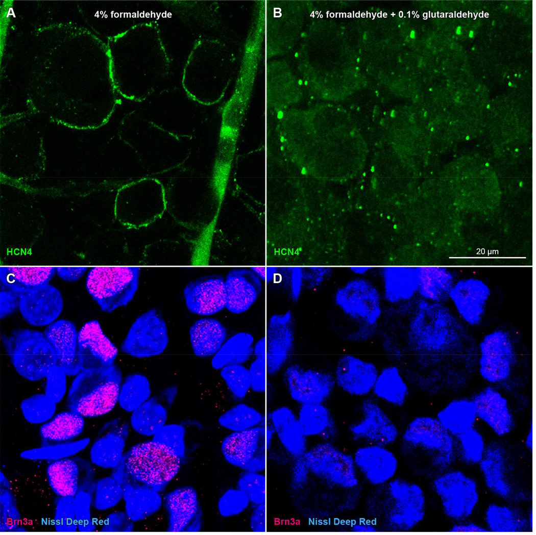
Figure 4.
Immunoreactivity loss in rat retinae in glutaraldehyde-containing fixative. Panels show single optical sections through the ganglion cell layer of the retinae of a single rat, one fixed in formaldehyde (A, C) and the other fixed in a mixture of formaldehyde and glutaraldehyde (B, D). Whole-mounted retinae were incubated in (A–B) mouse monoclonal anti-HCN4 antibody and DyLight 488-conjugated anti-mouse secondary antibody (green), or (C–D) goat anti-Brn3a antibody and DyLight 549-conjugated anti-goat secondary antibody (red), followed by NeuroTrace deep-red fluorescent Nissl stain (blue). Scale bar in (B) is 20 µm and applies to (A–D).

