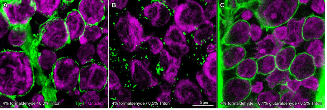
Figure 5.

Glutaraldehyde fixation protects against detergent-induced aggregation of Thy1 in dark-adapted rat retinae. Whole-mounted retinae incubated in anti-Thy1 primary antibody and Alexa Fluor 488-conjugated anti-mouse secondary antibody (green), and imaged on a laser scanning confocal microscope. Nuclei counsterstained with Qnuclear Deep Red Stain (magenta). Panels show single optical sections through the ganglion cell layer of retina fixed in formaldehyde (A, B) or a mixture of formaldehyde and glutaraldehyde (C). Retinae in A–C from same animal (A, B from one eye; C from the opposite eye) and processed in parallel. Staining pattern in formaldehyde-fixed tissue circumscribes cell profiles (A). Treatment of formaldehyde-fixed tissue with Triton-X 100 results in a discontinuous punctate staining pattern (B). Addition of glutaraldehyde to fixative prevents the detergent-induced change in Thy1 staining (C). Scale bar in (B) is 10 µm and applies to (A–C).
