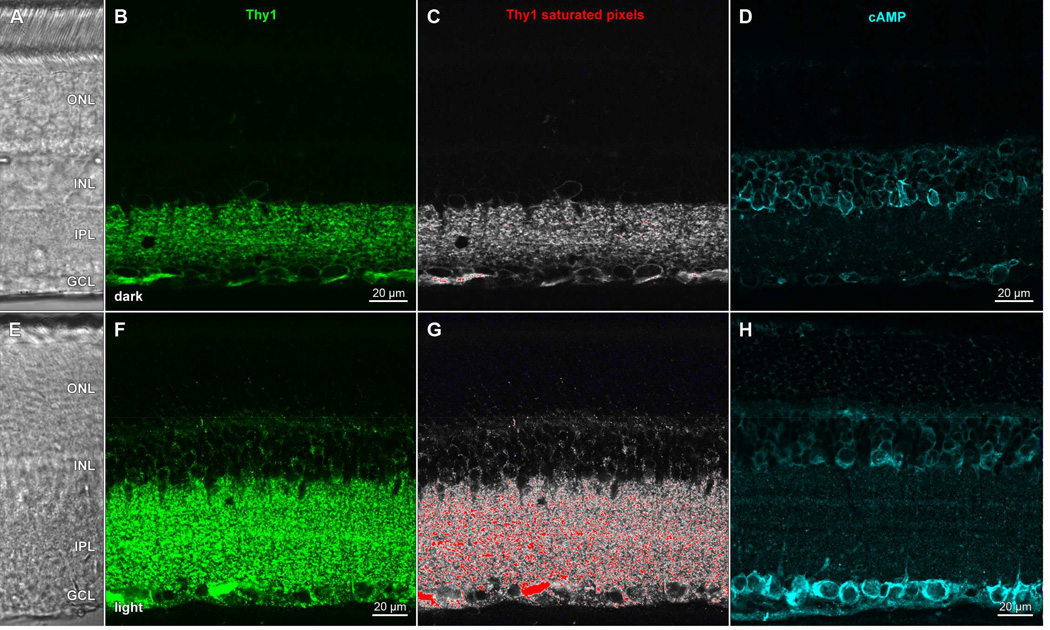
Figure 6.
Light exposure and detergent treatment induce a change in Thy1 staining pattern and an elevation in cAMP levels in rat retina. Transretinal vibratome sections of dark- (AD) and light- (E–H) adapted retinae collected, fixed in a mixture of formaldehyde and glutaraldehyde, and processed side-by-side, as in Ogata et al., (2012); incubated in (B,F) anti-Thy1 primary antibody and Alexa Fluor 488-conjugated anti-mouse secondary antibody (green), or (D,H) anti-cAMP primary antibody and DyLight 549-conjugated antimouse secondary antibody (cyan). All staining buffers contained Triton-X 100 detergent. Panels show single optical sections (B,D,F,H) of fields under epifluorescence illumination imaged at the same settings (laser intensity, photomultiplier gain, pinhole diameter) on a laser scanning confocal microscope, or fields imaged under differential interference contrast optics (A,E). Thy1 data from (B,F) visualized with a pseudocolor table in which saturated pixels are assigned a red color (C, G). Acronyms positioned at retinal layers as in Fig. 1. Scale bar in (B) is 20 µm and applies to (A–C). Scale bar in F is 20 µm and applies to (E–G).

