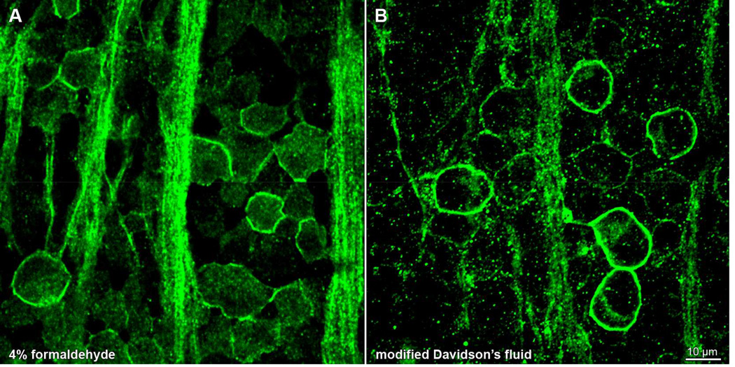
Figure 7.
Fixative type gives a differential staining pattern of HCN4 antibody in rat retina. Whole-mounted retinae (from opposite eyes of same animal) fixed in (A) 4% formaldehyde or (B) modified Davidson’s fluid, processed in parallel, and stained with anti-HCN4 primary antibody and Alexa Fluor 488-conjugated anti-mouse secondary antibody (green). Each panel shows a single optical section through the ganglion cell layer, collected on a laser scanning confocal microscope. Scale bar in (B) is 10 µm and applies to both panels.

