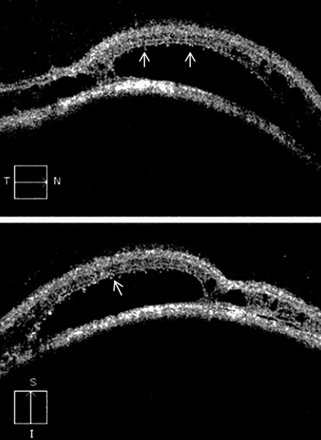
Figure 2.
Spectral domain optical coherence tomography, of the retina, over the tumour mass shows a dome-shaped retinal elevation with hyper-reflective retinal pigment epithelium. Neurosensory detachment with splitting of the retinal layers, at some places, is observed. Large cystoid spaces are noticed that are localised in the outer plexiform layer. These cystoid spaces are not of uniform thickness. There are multiple bridging strands crossing in the cystoid spaces. Occasional tiny pockets of cystoid changes are seen in the inner retina. In areas of neurosensory detachment, outer photoreceptor layer shows the presence of granularity (arrow).

