Figure 1.

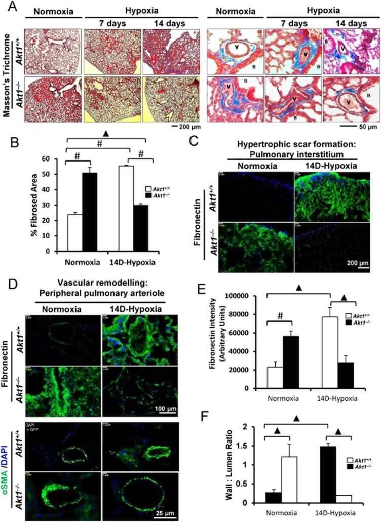
Akt1 deficiency protects against hypoxia-induced pulmonary remodelling. (A) Masson's trichrome stained section of the pulmonary interstitium (left) and peripheral pulmonary arterioles (right) of Akt1+/+ and Akt1−/− mice subjected to normoxia or chronic hypoxia for 7 and 14 days (n = 3–5 mice/group). (B) Histogram showing quantification of the fibrosed area in Akt1+/+ and Akt1−/− mice lungs after 14 day hypoxia compared with normoxia. (C) Immunostaining of frozen sections of 14 day hypoxia and normoxia Akt1+/+ and Akt1−/− mice lungs showing fibronectin expression in the interstitium. (D) Fibronectin and αSMA immunofluorescence staining in and around small pulmonary arteries of normoxic and 14d-hypoxic Akt1+/+ and Akt1−/− mice. (E) Histogram showing reduced fibronectin expression in Akt1−/− mice hypoxic lung sections compared with Akt1+/+ mice lungs (n = 4–5 mice/group). (F) Histogram showing vascular wall to lumen ratio in Akt1+/+ and Akt1−/− mice lung hypoxic sections measured from αSMA immunofluorescence (n = 3–5 mice/group). V, vasculature; B, bronchiole. #P < 0.01, ▲P < 0.001.
