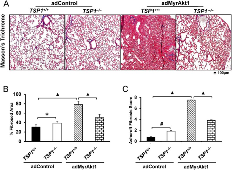
Figure 8.

TSP1−/− mice are protected from adMyrAkt1-induced pulmonary fibrosis. (A) Masson's trichrome staining of WT and TSP1−/− mouse lung harvested 14 days after i.t. adenovirus gene transfer of control vector or adMyrAkt1 (constitutive active Akt1) (n = 3 mice/group). (B and C) Per cent fibrosed area quantified using ImageJ software, and Ashcroft fibrosis score of the degree of fibrosis in WT and TSP1−/− mice subjected to control vector or adMyr-Akt1 respectively. *P < 0.05, #P < 0.01, ▲P < 0.001.
