
FIGURE 1.
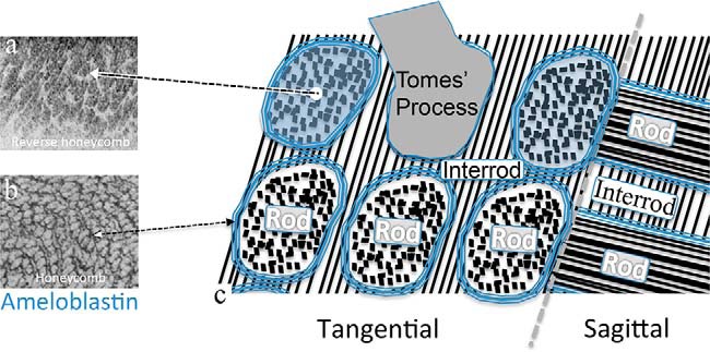
Idealized schematic of cellular fabrication showing the organization of mouse enamel extracellular matrix with HAP crystallite bundles within enamel rod-to-interrod microstructure. a shows developing porcine enamel with newly secreted ameloblastin localized to all of the area of the Tomes' processes (43), but it soon becomes localized exclusively to the perimeter of each enamel rod as a sheath as shown in b (40, 45). c, is a tangential (en face) and sagittal section schematic view of forming mouse enamel. The boundary (sheath) of each enamel rod corresponds to the lateral limit of each ameloblast cell's Tomes' process. The enamel rod is the basic unit of enamel and is created by a single ameloblast cell. Within a rod, the crystallites are packed parallel to one another along their long axis. The interrod crystallites form within the matrix deposited among adjacent ameloblasts, thereby giving rise to the continuity of the highly patterned rod-to-interrod enamel microstructure (22).