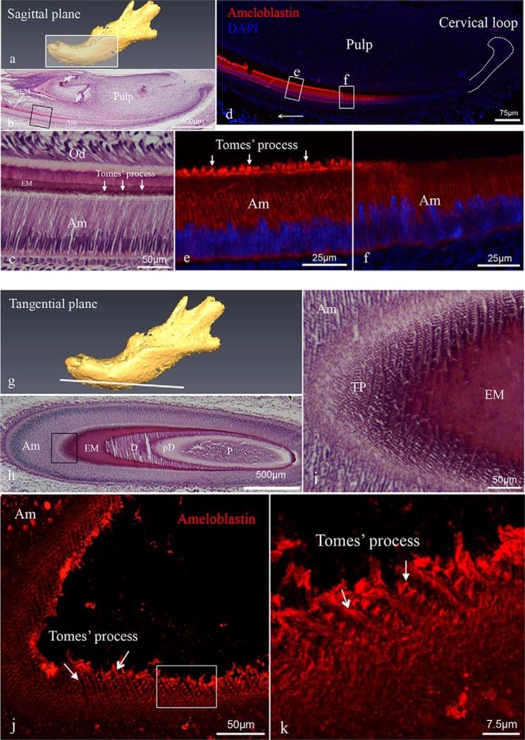
FIGURE 2.
Histology of mouse incisors and immunolocalization of ameloblastin in incisors. Sagittal (a–f) and tangential (g–k) sections were prepared from PN3 mouse mandibles. a is a schematic diagram of a tissue section of the jaw cut on the sagittal plane. b and c are hematoxylin-eosin-stained sagittal sections showing a mandibular incisor with c being a higher magnification of the boxed area in b, and arrows identifying Tomes' processes. d–f show representative immunofluorescent staining of ameloblastin (red fluorochrome) in sagittal sections with DAPI staining used for nuclear localization. Immunoreactivity showed increasing intensity in the area corresponding to the secretory stage ameloblast cells (e) relative to the pre-secretory stage cells (f). e and f are higher magnification views of the corresponding boxed areas in d. Arrows in e indicate intense immunostaining in the Tomes' processes. g is a schematic diagram of a tissue section of the jaw cut on the tangential section. h and i are hematoxylin-eosin-stained tangential sections showing a mandibular incisor with i being a higher magnification view of the boxed area in h. j and k are three-dimensional reconstructions of confocal Z-stack images from tangential sections, in which the immunodetection signal reveals ameloblastin antigen distributed across each Tomes' process (arrows), forming a decussating pattern of staining, which corresponds to the rod arrangement in mature mouse enamel. k is a higher magnification view of the area boxed in i. The abbreviations used are as follows: Am, ameloblast; EM, enamel matrix; D, dentine; pD, pre-dentine; P, pulp; Od, odontoblast; TP, Tomes' process.

