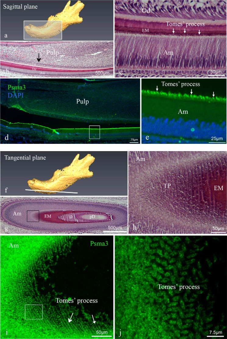
FIGURE 3.
Histology of mouse incisors and immunolocalization of Psma3 in incisors. Sagittal (a–e) and tangential (f–j) sections were prepared from PN3 mouse mandibles. a is a schematic diagram of a sagittal section through the jaw. b and c are hematoxylin-eosin-stained sagittal sections showing a mandibular incisor with c being a higher magnification view of the approximate area in b shown by the black arrow. Arrows in c identify the Tomes' processes. d and e show representative immunofluorescent staining for Psma3 (green fluorochrome) in a sagittal section with DAPI staining used for nuclear localization. Psma3 was detected in the cytoplasm of ameloblasts and Tomes' processes. The higher magnification image in e shows that intense immunoreactivity was observed in the distal membrane projections of the secretory ameloblasts, i.e. Tomes' processes (arrows). f is a schematic diagram of the plane from a tangential section of the jaw. g and h are a hematoxylin-eosin-stained tangential section showing a mandibular incisor, with h being a higher magnification view of the boxed area in g. i and j are three-dimensional reconstructions of confocal Z-stack images of immunostaining of Psma3 from tangential section. j is a higher magnification view of the approximate area boxed in i. In these reconstruction images, Psma3 was detected and localized to each of the Tomes' processes (arrows), producing a decussating pattern consistent with the arrangement of rods in mature mouse enamel. The abbreviations used are as follows: Am, ameloblast; EM, enamel matrix; D, dentine; pD, pre-dentine; Od, odontoblast; TP, Tomes' process.

