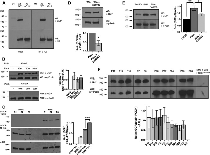
FIGURE 3.
In vivo phosphorylation of γ-Pcdh Ser-922 by PKC. A, analysis of HEK293 cells transfected with A3-WT or A3-ΔC15 shows that anti-GCP recognizes the C-terminal motif (input, left). Immunoprecipitation (IP) for HA tag confirms that the appropriately sized band recognized by anti-GCP is in fact γ-Pcdh-A3 (right). WB, Western blot; UT, untransfected. B, HEK293 cells transfected with A3-WT or A3-S/A and incubated with 648 nm PMA for the indicated times. The anti-GCP signal increases after PMA treatment but not when Ser-922 is mutated to a non-phosphorylatable alanine. The graph shows quantification of three experiments ± S.E. with A3-WT. No significant differences between the three PMA time points were found. A.U., arbitrary units. C, comparison of short and long film exposures of vehicle (DMSO) and PMA-treated HEK293 cells transfected with A3-WT (3 replicates (R)). The graph shows quantification of this blot, confirming a significant increase in anti-GCP signal with PMA treatment. Note that endogenous phosphorylation is observed at longer exposures (A and C). D and E, Gö6983 and PMA exposure in HEK293 (D) and cortical neuron cultures (E). Anti-GCP signal increases with PMA, and this increase is significantly blocked by treatment with PKC inhibitor Gö6983 (1 μm). The graphs show the mean ± S.E. of three experiments. F, anti-GCP (upper blots) detects a portion of endogenous γ-Pcdhs (all γ-Pcdhs were detected with mAb N159/5, lower blots) in embryonic brain and postnatal cerebral cortex. The lack of signal in Emx1-Cre;Pcdh-γfcon3/fcon3 cortically restricted mutants (9) confirms antibody specificity. Although the proportion of phosphorylated γ-Pcdhs trends lower at later postnatal ages, no significant age differences were found (graph shows results of three experiments ± S.E.). *, p < 0.05; **, p < 0.01; ***, p < 0.001.

