
FIGURE 2.
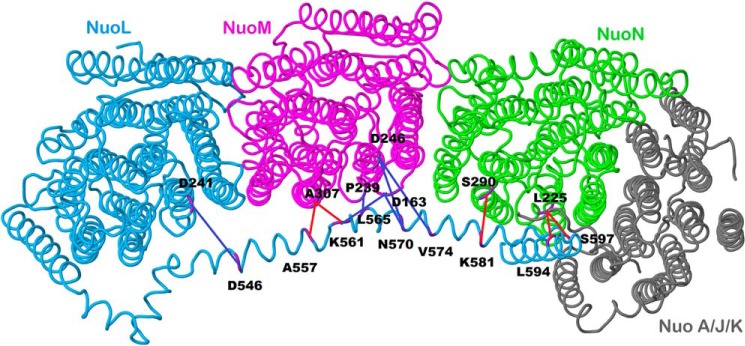
Schematic view of the membrane arm of Complex I showing the sites of cross-links. Subunit L is colored blue, subunit M is magenta, subunit N is green, and subunits A, J, and K are colored gray. Short distance cross-links, which were formed by disulfide bonds, are colored red, whereas longer distance cross-links formed by bifunctional methane thiosulfonates are colored blue. The image was developed from Protein Data Bank code 3rko (16). The view is from the cytoplasm. The peripheral arm would be to the right.